

Retinal detachment
Early diagnosis and surgery performed by expert hands are the two main factors in ensuring the best outcome.
What is Retinal Detachment?
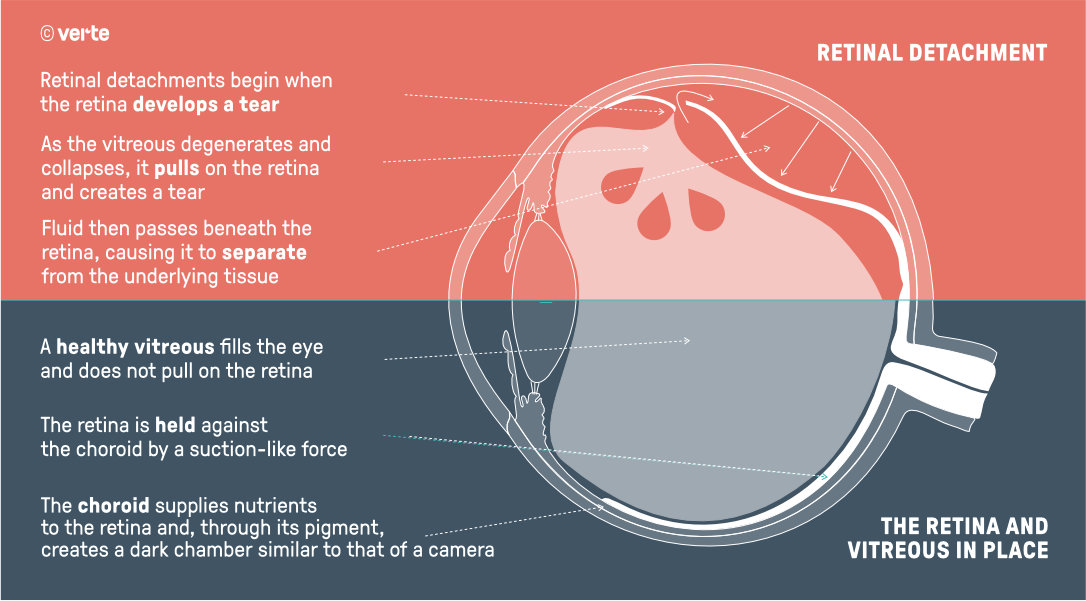
The retina is the innermost layer of the posterior half of the eye. A retinal detachment is the separation of the retina from the next layer, called the choroid.
It is a serious visual problem that can occur at any age, although it usually occurs in middle-aged or elderly individuals. It tends to be more frequent in people who are short-sighted or in those who have previously had some disorder of the retina.
Retinal detachment affects one in every 10,000 people per year.
What causes it?
The majority of retinal detachments are caused by the presence of one or more tears in the structure of the retina.
Inside the eye, in the cavity defined between the crystalline lens and the retina, lies the vitreous body. The vitreous is a transparent gel that is slightly adherent to the retina over most of its surface but firmly adherent at certain specific points. Traction of the vitreous at these points can cause a tear or hole in the retina.
The vitreous also suffers the passage of time. Its ultrastructure is altered and its capacity to retain water is reduced, causing it to lose volume. As it can no longer occupy all the interior space it should, it must separate from the retina: this is known as Posterior Vitreous Detachment. The majority of vitreous detachments occur naturally with ageing in healthy eyes and do not cause damage to the retina. However, in some patients retinal tears do appear. In other cases, such as in myopia, an inflammatory process or accidents involving ocular trauma, vitreous detachment can occur in a more dangerous way.
In these cases, a tear first occurs in the retina, through which the fluid from the vitreous humour penetrates beneath the photoreceptor layer, facilitating the separation of the retina and the subsequent detachment. The area of retina that has detached cannot function correctly and will produce an area of blurred vision or blindness.
Occasionally, vitreous detachment causes the rupture of a retinal vessel and the spillage of blood into the vitreous cavity: this is known as vitreous haemorrhage.
Some retinal detachments are caused by other eye diseases, such as tumours, severe inflammations or complications of diabetes. These are known as secondary retinal detachments, in which tears or holes in the retina do not always exist.
What symptoms does retinal detachment produce?
The most characteristic onset is the perception of floating black dots, commonly known as "floaters" (myodesopsia) and flashes of light in the lateral vision (photopsia).
In other cases, patients may directly notice an undulation or veil in their vision or the appearance of a shadow in a lateral area of the visual field (dark curtain). The development of a retinal detachment will reach central vision and create a significant loss of vision if it is not treated quickly and effectively.
Although many people think otherwise: the retina does not hurt. Thus, all these processes present only with visual symptoms, without pain or discomfort.
How is retinal detachment diagnosed?
A detached retina cannot be seen from outside the eye. Consequently, if symptoms are noticed, an ophthalmologist should be consulted as soon as possible. The ophthalmologist examines the retina and the rest of the internal structures of the eye in a painless and comprehensive manner, using an instrument called an ophthalmoscope. Other special diagnostic instruments that may be used include special contact lenses, slit lamp, ultrasound and optical coherence tomography (OCT).
How is retinal detachment treated?
There are no eye drops or other pharmacological treatments that cure retinal detachment.
In an early stage, if the retina is torn and retinal detachment has not yet begun, it can be prevented by early laser treatment applied in the clinic. This is known as retinal photocoagulation. Once the retina has detached, the only treatment is a series of surgical techniques that reattach the retina.
The surgical procedures for retinal diseases must be performed by Retinal Surgeons, ophthalmologists specialised in the retina with sufficient surgical training. It is a complex subspecialty due to the wide variety of cases and the sophistication of new instruments and operative techniques.
How can retinal detachment be prevented?
It is important for people with myopia or with family members who have had retinal detachment to have regular eye examinations by a Retinal Specialist so that early detection of changes in the vitreous or retina can be achieved, and peripheral predisposing retinal lesions can be treated with argon laser photocoagulation.
It is also important to examine the contralateral eye in those people who have already suffered retinal detachment in one eye.
Predisposing lesions are alterations observed in the peripheral retina about which a statistical relationship with retinal tears leading to detachment is known. The retinal specialist has the greatest knowledge of the degree of risk of each of the predisposing lesions in order to decide on the suitability of preventive laser treatment.
The sudden appearance of floaters (myodesopsia) or a sudden increase in existing ones, as well as the appearance of flashes of light, should be quickly consulted with your Ophthalmologist or with an Ophthalmic Emergency Service.
Can retinal detachment lead to blindness?
More than 90% of retinal detachments can be cured, the majority in a single operation, but some require several interventions. Thanks to modern microsurgical techniques and the experience of Retinal Surgeons, fewer and fewer cases cannot be resolved. Unfortunately these eyes may lose their vision completely.