Glaucoma: diagnosis and treatment in Barcelona
The danger of gradually losing your visual field without realising it for a long time: the silent blindness.
What is glaucoma?
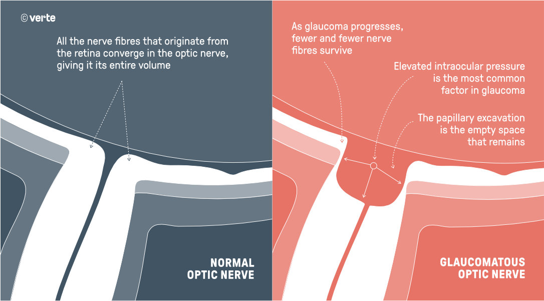
Glaucoma is a disease of the optic nerve (neuropathy) that causes progressive vision loss. It is usually caused by a silent rise in intraocular pressure (IOP), although it is also a multifactorial disease: factors such as high myopia, sleep disorders with apnoea or insufficient blood supply to the optic nerve can all contribute to its deterioration.
To understand what happens inside the eye, Dr Susana Duch, director of our Glaucoma Unit, explains it with a simple image:
"Imagine the eye as a water circuit, with an inlet and an outlet that maintain a balance. When that circuit ages and stops working properly, the pressure rises gradually. Even though you do not notice it, that pressure is damaging the optic nerve fibres, one by one. By the time you start to realise it, many have already been lost."
The damage caused by glaucoma is irreversible, but with early diagnosis and appropriate treatment we can halt its progression and preserve vision for many years.
The "silent blindness": why early detection matters
Chronic glaucoma is the second leading cause of irreversible blindness in the world, after cataracts. But unlike other serious diseases, glaucoma gives no warning: it progresses for years without symptoms, without pain and without the patient noticing any vision loss until it is too late.
This is why it is called "the silent blindness": half of the people who have glaucoma do not know they have it.
The good news: up to 95% of cases could be prevented with appropriate and early diagnosis. Early detection is the best tool we have today against this disease.
Symptoms: how does chronic glaucoma present?
Chronic glaucoma is insidious because it produces no perceptible symptoms in its early stages. Only when the disease is advanced does the patient begin to notice signs:
- Progressive loss of peripheral visual field — initially in small areas, generally without the patient noticing because the brain compensates.
- Difficulty adapting to sudden changes in light — when moving from a bright environment to a dark one (or vice versa), the glaucoma patient may feel "blinded" for a few seconds.
- Impairment of central vision — only in very advanced stages, when most peripheral vision has already been lost.
It is important not to confuse chronic glaucoma with acute glaucoma, an ophthalmic emergency with very different symptoms (intense pain, red eye, blurred vision, nausea). If you experience these symptoms, contact our 24-hour Ophthalmic Emergency Service immediately.
Causes and risk factors
Chronic glaucoma is a multifactorial disease: there are several factors that increase the risk of developing it.
- Elevated intraocular pressure (IOP). This is the main risk factor and, to date, the only one that is truly treatable. Normal IOP ranges between 10 and 20 mmHg.
- Direct family history. Glaucoma has a significant genetic component. If a first-degree relative (parents, siblings, children) has glaucoma, your risk is multiplied.
- Myopia greater than 6 dioptres. Highly myopic eyes have a more vulnerable optic nerve. Furthermore, diagnosis is more difficult because visual field loss can be confused with myopia itself.
- Age over 45 years. Prevalence increases progressively with age.
- African descent. Glaucoma is particularly aggressive in patients of Black ethnicity, with an earlier onset and faster progression.
- Long-term corticosteroid treatment. 30–40% of the population experiences a rise in IOP in response to corticosteroids (particularly eye drops or injections).
- Sleep apnoea, nocturnal hypotension. Vascular factors that affect blood supply to the optic nerve.
To explore these factors further, we recommend reading our content on glaucoma and high myopia and on medications that can cause glaucoma.
Early diagnosis: how we detect glaucoma at VERTE
Glaucoma diagnosis is not made by measuring intraocular pressure alone. IOP is the main risk factor, but glaucoma is a disease of the optic nerve, and therefore only by evaluating the state of the nerve (its structure and function) can we confirm the diagnosis.
This is why at VERTE we have comprehensive and differentiated diagnostic equipment that allows us to detect glaucoma even before vision loss occurs:
Structural technology
- 3 OCT devices (Optical Coherence Tomography) — we have the Zeiss Cirrus, Topcon Triton and Yalkaid TowarPi. This diversity allows us to cross-reference measurements and increase diagnostic reliability.
- Zeiss Forum system — analyses the longitudinal progression of your tests over time using regression curves, detecting real glaucoma progressions that other systems might miss.
Functional technology
- Humphrey Field Analyzer 3 (HFA3) perimeter — using the SITA Faster 24-2c strategy, currently the most advanced for evaluating the visual field.
Intraocular pressure measurement
At VERTE we do not rely on a single tonometer. We have 5 complementary tonometry methods, allowing us to obtain a true IOP corrected for the characteristics of each cornea:
- Goldmann — the historical gold standard.
- Pascal DCT — independent of corneal thickness.
- CORVIS — the only tonometer that measures IOP corrected for corneal biomechanics, a technology that most clinics do not have.
- Perkins — for examination with the patient in a supine position.
- Tonopen — for special situations.
Corneal assessment
- Pachymetry with Pentacam Corvis — measures corneal thickness and biomechanics to adjust the interpretation of IOP.
Glaucoma treatment: 3 approaches depending on your case
Glaucoma cannot be cured, but it can be controlled. The aim of treatment is to lower IOP to the appropriate level for your optic nerve, to halt the damage and preserve your vision. We offer three complementary therapeutic approaches that we apply on an individualised basis:
Medical treatment
Medical treatment of glaucoma is the first line in most cases. It is based on hypotensive eye drops (beta-blockers, prostaglandin analogues, brimonidine, carbonic anhydrase inhibitors...). The main challenge is patient adherence and tolerance to side effects.
Laser treatment
Selective Laser Trabeculoplasty (SLT) is an outpatient procedure, without incision, that reduces IOP by 20–30% in approximately 70% of patients. It is now a first-line option in many cases, according to recent evidence.
Surgical treatment
When IOP cannot be controlled with eye drops or laser, we turn to surgery. We offer the full range of techniques: MIGS (minimally invasive surgery with iStent, Hydrus, XEN implants), trabeculectomy, valved implants (Ahmed) and non-valved implants (Baerveldt, Paul), and our own Piggy-back technique (two implants without opening the eye).
Prevention: when and how to have a check-up
Glaucoma prevention is based on one thing: detecting those at risk in time. There is no other way. These are the intervals we recommend according to your profile:
- No risk factors, up to the age of 40. A check-up every 2 years.
- From the age of 40, with no risk factors. Annual check-up with IOP measurement and fundus examination.
- Direct family history of glaucoma. Annual check-up from the age of 35.
- Myopia greater than 6 dioptres. Annual check-up regardless of age.
- Long-term corticosteroid treatment. IOP monitoring every 3–6 months as medically indicated.
- Established glaucoma diagnosis. Individualised follow-up (every 3 to 12 months depending on severity).
IOP Express: quick intraocular pressure check
At VERTE we offer a unique service in Barcelona: the IOP Express, a quick intraocular pressure measurement with no prior appointment needed, within clinic opening hours. It is designed for patients already diagnosed who need a one-off check between appointments, or for people who want a quick initial screening with no commitment.
Glaucoma Unit in Barcelona
At VERTE, glaucoma is not managed by a single ophthalmologist: we have the largest Glaucoma Unit in Barcelona, with 8 glaucoma specialists dedicated exclusively to this condition.
Dr Susana Duch — Director of the Glaucoma Unit

The team is led by Dr Susana Duch, a national and international authority on glaucoma with over 35 years of experience in drainage implant surgery. She introduced the first drainage implants in Spain in 1990 (the Molteno valve) and trained the first Spanish users at congresses and in our own operating theatres and clinics. Member of SEG, EGS, SEO, SCO and AAO (American Academy of Ophthalmology). Her unit has treated thousands of glaucoma patients at all stages, from early diagnosis to the most complex cases requiring advanced surgery. A career that places her among the most influential glaucoma surgeons in Spain.
Dr Elena Millà — Glaucoma Specialist

Dr Carlos Arciniegas — Glaucoma Specialist
Glaucoma specialist, Dr Carlos Arciniegas offers specific dedication to microincisional surgery and drainage implants. European Fellowship in Ophthalmology (FEBO), Master's in Anterior Segment from the Institut Barraquer and Master's in Molecular Ophthalmobiology from the University of Valencia. Surgical trainer and EU consultant for the XEN implant (one of the most advanced glaucoma drainage devices available) between 2014 and 2015. Speaker at leading international congresses including ASCRS and EGS.
The team is completed by Dr Elena Ávila, Dr Naiara Relaño, Dr Shirin Djavanmardi, Dr Núria Gabarró and Dr Gloria Segura.
VERTE is also a training centre: over the past two decades, a significant number of glaucoma surgeons from other national and international centres have received surgical and clinical training at our facilities under the direction of Dr Duch.
Our own research: our science underpins our practice
Unlike most ophthalmology clinics, the VERTE team does not merely apply science: it produces it. Our doctors have been signing scientific publications in international journals for over two decades, and VERTE patients benefit directly from that in-house research.
Some representative publications from our team:
- Duch et al. (2001) — IOP after refractive surgery and corneal thickness.
- Milla et al. (2009) — Pascal tonometry versus corneal biomechanics.
- Buchacra et al. (2011) — Documentation of the first iStent implanted in Spain, at VERTE.
- Millà et al. (2013, 2017, 2024) — Studies on the genetics of glaucoma.
- S. Duch et al. (2021) — Drainage implants in refractory glaucoma.
- Oliver-Gutiérrez et al. (2025) — Comparative study of Paul vs Baerveldt in complex glaucomas.
- Robles Amor et al. (2025) — Glaucoma in patients with high myopia.
- Oliver-Gutiérrez et al. (2025) — SLT as a first-line treatment.
This track record places us among the reference centres for glaucoma in Europe.
Comprehensive support for the glaucoma patient
Living with glaucoma is not just a matter of eye drops and check-ups. It is a chronic disease that also affects mood, quality of life and one's relationship with oneself. This is why at VERTE we have built a network of complementary services around the Glaucoma Unit:
- Psychological Counselling Service for Visual Loss — led by Clara Duch, it helps patients manage the emotional impact of diagnosis and develop coping strategies.
- Integrated Dry Eye Unit — long-term use of hypotensive eye drops can cause ocular surface toxicity. Our Dry Eye Unit treats these side effects to ensure the treatment remains sustainable in the long term.
- Telephone follow-up appointments — to resolve queries between check-ups without the need to travel.
- Weekly team clinical session — complex cases are discussed as a team every week, ensuring that no patient receives a decision based on a single opinion.
Acute glaucoma: a different emergency
Acute glaucoma is a completely different clinical entity from chronic glaucoma. While chronic glaucoma advances silently over years, acute glaucoma presents suddenly, with intense pain, red eye, blurred vision and sometimes nausea and vomiting. It is an ophthalmic emergency requiring immediate attention.
Book your glaucoma check-up at VERTE Barcelona
Prevention is the best option. If you are over 40, have a family history of glaucoma, high myopia or any other risk factor, do not wait. A timely check-up can make the difference between preserving your vision or not.