

Epiretinal membrane
One of the most common retinal conditions, with an excellent microsurgical solution in experienced hands.
What is the macular epiretinal membrane?
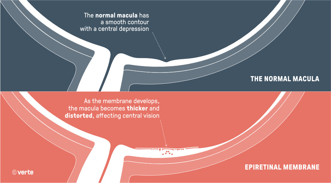
The epiretinal membrane (meaning "membrane over the retina") is an ocular condition consisting of the development of a translucent cellular membrane over the central area of the retina (macula), which is the fundamental area for vision.
When this membrane contracts and wrinkles, it causes progressive deformation of the retina with consequent visual disturbance. The most frequent symptom is loss of visual acuity and distortion of images (also called metamorphopsia).
Is this condition very common?
These epimacular membranes affect 2% of the population from the age of 50. Initially they present as microfolds and a subtle sheen over the macula (cellophane maculopathy), which can make them difficult to detect. These membranes tend to progress slowly and appear in up to 20% of people by the age of 75. In many cases they do not progress and produce no obvious symptoms, making their removal unnecessary.
What causes it?
The majority of epiretinal membranes in people over 60 are considered "idiopathic", meaning of unknown cause. However, it is currently believed that their origin lies in the ageing process undergone by the gel that fills the interior of the eye (vitreous humour), which over the years ends up separating from the retina (posterior vitreous detachment). In some people this process releases glial cells (cells responsible for retinal repair), which trigger the formation of the membrane.
It may also be associated with trauma, vascular conditions (diabetes or retinal venous thrombosis), following vitreoretinal surgery or as a complication of a retinal detachment.
What symptoms does the macular epiretinal membrane cause?
Initially, the patient experiences mild distortion of images, wavy lines and alterations in written text (smaller letters, numbers, etc.). When the traction exerted by the epiretinal membrane on the macula increases, reading deteriorates. It is also common for the patient to notice blurred vision in the central visual area. Metamorphopsia is confirmed with the Amsler grid test and blurred vision with the visual acuity optotype projection.
How is it diagnosed?
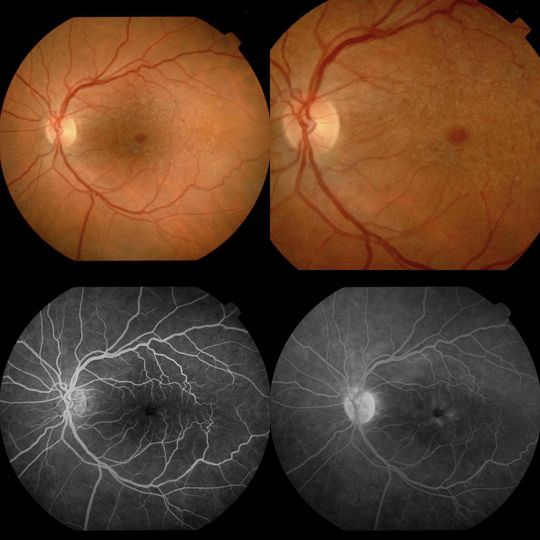
To detect a macular epiretinal membrane, the ophthalmologist examines the fundus of the eye during the consultation. The best complementary test is Optical Coherence Tomography (OCT), although it is sometimes advisable to add fluorescein angiography (FA). Assessment of the images obtained with this test allows the ophthalmologist to quantify and measure the retinal structures, determine their alteration and plan the treatment.
When should it be treated?
In milder cases, watchful waiting with periodic monitoring may be chosen, without the need for surgery. However, when vision deteriorates, images become distorted and macular anatomy worsens, vitreoretinal surgery (posterior vitrectomy) should be considered.
Is surgical intervention effective?
This is one of the retinal surgeries with the best anatomical and visual outcomes. Vision lost during the membrane growth process can be recovered, but on average only half of what was lost is regained. For this reason, it is not advisable to allow vision to deteriorate too much before intervening.
Can the condition recur after surgery?
The recurrence rate (regrowth of the membrane) for this type of lesion treated by vitrectomy is less than 5% of cases, being more frequent in younger patients.
Can it lead to blindness?
The progression of this condition without treatment leads to significant loss of central vision. Vision is not completely lost because it only affects the macula and spares the rest of the retina, although the final result of central vision loss can be very debilitating. Surgical intervention is therefore generally recommended in a timely manner to halt the process and, as far as possible, reverse it.